Understanding conjunctivitis in companion animal | Dechra UK

Conjunctivitis

Anatomy and Physiology of the Conjunctiva

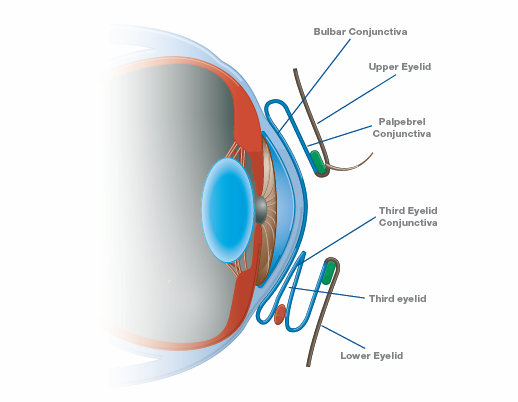
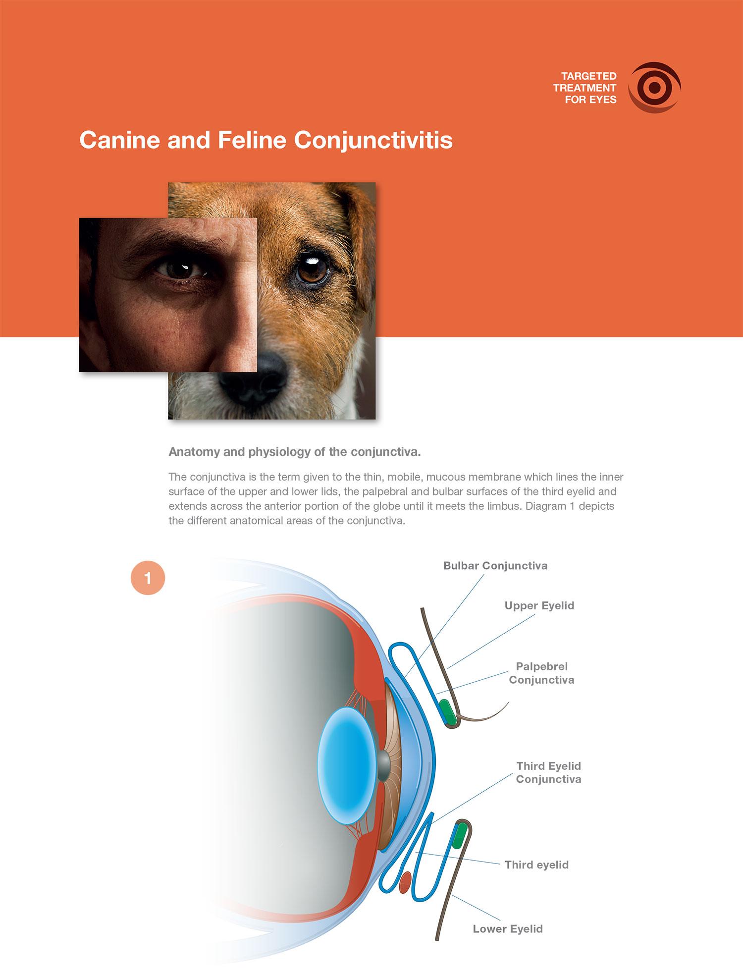
The conjunctiva is the term given to the thin, mobile, mucous membrane which lines the inner surface of the upper and lower lids, the palpebral inner and bulbal surfaces of the third eyelid and the anterior portion of the globe (until it meets the limbus). Diagram 1 depicts the different anatomical areas of the conjunctiva.

Conjunctivitis diagram

The conjunctiva consists primarily of stroma, which is split into a deeper fibrous layer (mainly connective tissue, blood vessels and nerves) and a more superficial adenoid layer, which contains conjunctiva-associated lymphoid tissue.

Above this sits the conjunctival epithelium, which contains goblets cells. The conjunctival epithelium is coated by the pre-ocular tear film, which provides the conjunctiva with protection and nutrition.

Diagram 1 - Anatomy of the conjunctiva

Given the close association between the tear film and the conjunctiva, any abnormality of either will impact the other. The goblet cells within the conjunctival epithelium are responsible for the production of the mucin component of the tear film, therefore a reduction in these cells can cause reduced tear film stability and therefore secondary corneal disease. Equally, if there is a change in the tear film, it has the potential to impact on conjunctival health, leading to conjunctival disease.

The conjunctiva is a highly exposed mucous membrane, and is highly responsive to noxious stimuli due to its rich vascular supply and lymphoid content. Clinically, it is useful to be able to distinguish between the superficial conjunctival vasculature, and that of the deeper episclera (hyperaemia of which may indicate the presence of sight-threatening disease as opposed to superficial surface ocular disease).

The conjunctival vessels are fine, freely mobile and frequently branching. They are pink to lighter red in appearance and often form looping patterns. When 2.5% phenylephrine is applied to the eye, these vessels should blanch easily. This can be contrasted to the deeper episcleral vessels which are wider, fixed in position and darker red in appearance. Each individual vessel can be seen more distinctly, branching only on occasion. Equally, they blanch much more slowly upon topical application of phenylephrine.

Just like skin, the conjunctiva have a ‘normal’ commensal bacterial population. Studies have reported that the majority of bacteria cultured from the conjunctiva of clinically normal cats and dogs are Gram-positive, with Staphylococcal spp being most common1,2. It is important to understand the normal flora when assessing potential pathology in small animal practice.

An overview of small animal conjunctivitis

Inflammation of the ocular mucous membranes, or conjunctivitis, is commonly encountered in small animal first opinion practice, and therefore you would assume that diagnosis and treatment would be easy. However, finding the root cause can sometimes be problematic.

The clinical signs of conjunctivitis include:

  1. conjunctival hyperaemia
  2. ocular discharge
  3. conjunctival oedema (otherwise known as chemosis)
  4. haemorrhage (both conjunctival and subconjunctival)
  5. conjunctival thickening / swelling / ulceration
  6. follicle formation
  7. pruritus

Unfortunately, these signs are very non-specific and can occur with other disease processes. Equally, given the reactive nature of the conjunctiva, it often becomes inflamed with almost all other ocular disease processes. Conjunctivitis can result in a red eye, however, not every red eye has conjunctivitis. It is essential that any eye with inflammation of the conjunctiva undergoes a full range of diagnostic testing to determine the cause. 

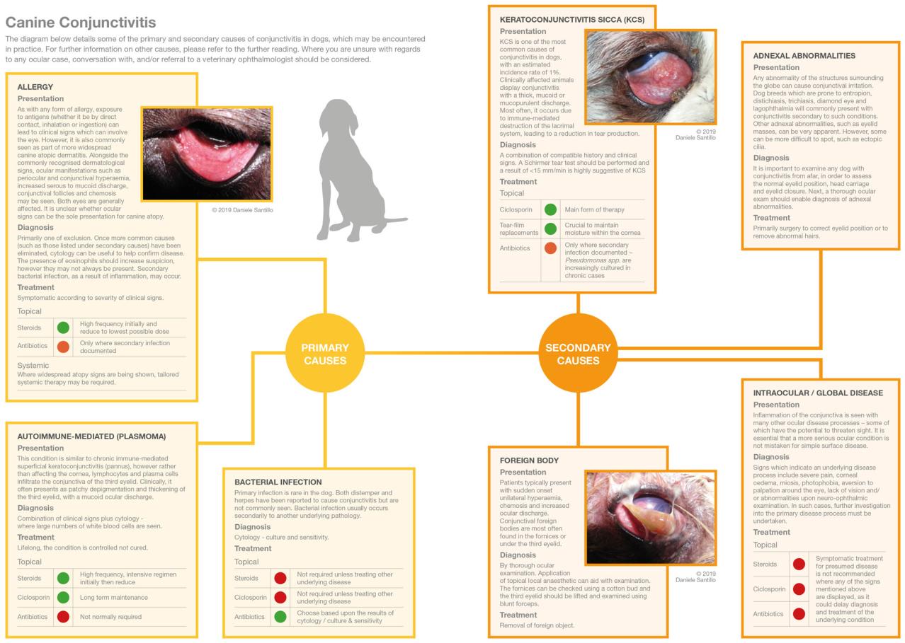
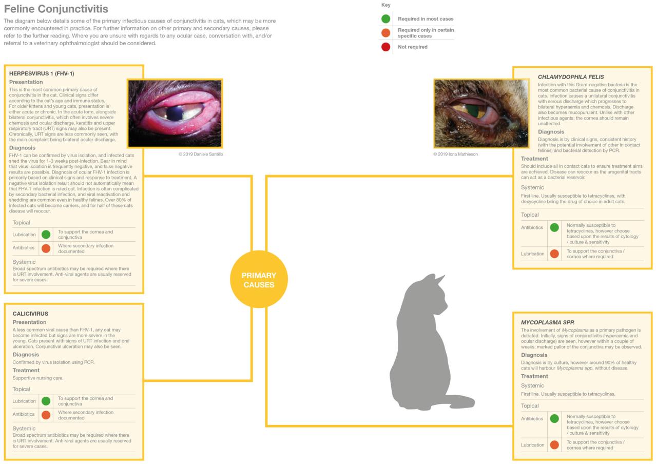
The most useful way to classify conjunctivitis would be by aetiology. There are multiple potential causes of conjunctival inflammation and these can be split into primary causes (including infection, allergic and environmental) and secondary (including uveitis, adnexal disease, trauma, foreign bodies and neoplasia).

In dogs, conjunctivitis is most commonly secondary in nature, as a result of adnexal abnormalities or as a result of keratoconjunctivitis sicca. In contrast for cats, primary infectious disease is more commonly encountered.

In some cases, it is not possible to classify disease by cause, therefore classification can based upon the duration of disease (e.g. acute, chronic, recurrent) or the appearance of any ocular discharge (e.g. mucoid, purulent, haemorrhagic).


Additional Support

The diagrams below details some of the primary and secondary causes of conjunctivitis in dogs and cats, which may be encountered. For further information on other causes, please refer to the references or further reading.

Where you are unsure with regards to any ocular case, conversation with, and/or referral to a veterinary ophthalmologist should be considered.

  1. Espinola MB and Lilenbaum W (1996) Prevalence of bacteria in the conjunctival sac and on the eyelid margin of clinically normal cats J Small Anim Pract 37: 364-366
  2. McDonald PJ and Watson DJ (1976) Microbial flora of normal canine conjunctivae. J Small Anim Pract 17(12): 809-812


Gelatt KN (Ed.) (1999) Veterinary Ophthalmology, 3rd ed. Lippincott Williams & Wilkins, Philadelphia

Heinrich C (2015) Assessing conjunctivitis in cats. Veterinary Times 45(20): 26-28

Heinrich C (2015) Assessing canine conjunctivitis. Veterinary Times 45(37): 28-32

Maggs DJ, Miller PE and Ofri R (Eds.) (2008) Slatter’s Fundamentals of Veterinary Ophthalmology, 4th ed. Saunders Elsevier, St. Louis, Missouri

Petersen-Jones S and Crispin S (Eds.) (2002) BSAVA Manual of Small Animal Ophthalmology. 2nd ed. British Small Animal Veterinary Association, Gloucester, UK

Oliver J (2012) Conjunctivitis in small animals: diagnosing and treating cases. Veterinary Times 42(16): 10-16

Turner S (2011) Focus on bacterial conjunctivitis: accurate diagnosis and treatment Veterinary Times 41(19): 32-33

keyboard_arrow_up